
Digestive systems
Digestive systems take many, many forms. There is a fundamental
distinction between internal and external digestion. External digestion
was the first to evolve, and most fungi still rely on it. In this process, enzymes are secreted into the environment surrounding the organism, where they break down an organic material, and some of the products diffuse back to the organism. Later, animals evolved
by rolling into a tube and acquiring internal digestion, which is more
efficient because more of the broken down products can be captured, and
the chemical environment can be more efficiently controlled.
Some organisms, including nearly all spiders, simply secrete biotoxins and digestive chemicals (e.g., enzymes)
into the extracellular environment prior to ingestion of the consequent
"soup". In others, once potential nutrients or food is inside the organism, digestion can be conducted to a vesicle
or a sac-like structure, through a tube, or through several specialized
organs aimed at making the absorption of nutrients more efficient.
Secretion systems
Bacteria use several systems to obtain nutrients from other organisms in the environments.
Channel transport system
In a channel transport system several proteins form a contiguous
channel traversing the inner and outer membranes of the bacteria. It is
a simple system, which consists of only three protein subunits: the ABC protein, membrane fusion protein (MFP), and outer membrane protein (OMP)[specify].
This secretion system transports various molecules, from ions, drugs,
to proteins of various sizes (20 - 900 kDa). The molecules secreted
vary in size from the small Escherichia coli peptide colicin V, (10 kDa) to the Pseudomonas fluorescens cell adhesion protein LapA of 900 kDa.
Molecular syringe
One molecular syringe is used through which a bacterium (e.g. certain types of Salmonella, Shigella, Yersinia) can inject proteins into eukaryotic cells. One such mechanism was first discovered in Y. pestis
and showed that toxins could be injected directly from the bacterial
cytoplasm into the cytoplasm of its host's cells rather than simply be
secreted into the extracellular medium.
Conjugation machinery

Schematic drawing of bacterial conjugation. Conjugation diagram 1- Donor cell produces pilus. 2- Pilus attaches to recipient cell, brings the two cells together. 3- The mobile plasmid is nicked and a single strand of DNA is then transferred to the recipient cell. 4- Both cells recircularize their plasmids, synthesize second strands, and reproduce pili; both cells are now viable donors.
The conjugation machinery of some bacteria (and archaeal flagella) is capable of transporting both DNA and proteins. It was discovered in Agrobacterium tumefaciens, which uses this system to introduce the Ti plasmid and proteins into the host which develops the crown gall (tumor). The VirB complex of Agrobacterium tumefaciens is the prototypic system.
The nitrogen fixing Rhizobia are an interesting case, wherein conjugative elements naturally engage in inter-kingdom conjugation. Such elements as the Agrobacterium
Ti or Ri plasmids contain elements that can transfer to plant cells.
Transferred genes enter the plant cell nucleus and effectively
transform the plant cells into factories for the production of opines, which the bacteria use as carbon and energy sources. Infected plant cells form crown gall or root tumors. The Ti and Ri plasmids are thus endosymbionts of the bacteria, which are in turn endosymbionts (or parasites) of the infected plant.
The Ti and Ri plasmids are themselves conjugative. Ti and Ri transfer between bacteria uses an independent system (the tra, or transfer, operon) from that for inter-kingdom transfer (the vir, or virulence, operon). Such transfer creates virulent strains from previously avirulent Agrobacteria.
Release of outer membrane vesicles
In addition to the use of the multiprotein complexes listed above,
Gram-negative bacteria possess another method for release of material:
the formation of outer membrane vesicles.[8]
Portions of the outer membrane pinch off, forming spherical structures
made of a lipid bilayer enclosing periplasmic materials. Vesicles from
a number of bacterial species have been found to contain virulence
factors, some have immunomodulatory effects, and some can directly
adhere to and intoxicate host cells. While release of vesicles has been
demonstrated as a general response to stress conditions, the process of
loading cargo proteins seems to be selective.
Phagosome
A phagosome is a vacuole formed around a particle absorbed by phagocytosis. The vacuole is formed by the fusion of the cell membrane around the particle. A phagosome is a cellular compartment in which pathogenic microorganisms can be killed and digested. Phagosomes fuse with lysosomes in their maturation process, forming phagolysosomes. In humans, Entamoeba histolytica can phagocytose red blood cells.
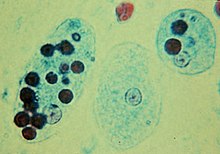
Trophozoites of Entamoeba histolytica with ingested erythrocytes
Gastrovascular cavity
The gastrovascular cavity
functions as a stomach in both digestion and the distribution of
nutrients to all parts of the body. Extracellular digestion takes place
within this central cavity which is lined with the gastrodermis, the
internal layer of epithelium.
This cavity has only one opening to the outside that functions as both
a mouth and an anus: waste and undigested matter is excreted through
the mouth/anus, which can be described as an incomplete gut.
Aboral end
Oral end
Mouth
Oral end
Aboral end
Exoderm
Gastroderm
Mesoglea
Digestive cavity
Medusa (left) and polyp (right)
In a plant such as the Venus Flytrap
that can make its own food through photosynthesis, it does not eat and
digest its prey for the traditional objectives of harvesting energy and
carbon, but mines prey primarily for essential nutrients (nitrogen and
phosphorus in particular) that are in short supply in its boggy, acidic
habitat.

Venus Flytrap (Dionaea muscipula) leaf
Specialized organs and behaviors
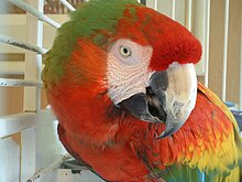
Catalina Macaw exhibits its seed shearing beak.

Squid beak and ruler for size comparison.

Teeth of a Carcharodon megalodon.

Rough illustration of a ruminant digestive system.
To aid in the digestion of their food animals evolved organs such as beaks, tongues, teeth, a crop, gizzard, and others.
Beaks
Macaws
primarily eat seeds, nuts, and fruit, using their impressive beaks to
open even the toughest seed. First they scratch a thin line with the
sharp point of the beak, then they shear the seed open with the sides
of the beak.
The mouth of the squid is equipped with a sharp horny beak mainly made of chitin[13]proteins.
It is used to kill and tear prey into manageable pieces. The beak is
very robust, but does not contain any minerals, unlike the teeth and
jaws of many other organisms, including marine species. The beak is the only indigestible part of the squid.
Tongue
Main article: Tongue
The tongue is skeletal muscle on the floor of the mouth that manipulates food for chewing (mastication) and swallowing (deglutition). It is sensitive and kept moist by saliva. The underside of the tongue is covered with a smooth mucous membrane. The tongue is utilised to roll food particles into a bolus before being transported down the esophagus through the use of peristalsis. The sublingual region underneath the front of the tongue is a location where the oral mucosa
is very thin, and underlain by a plexus of veins. This is an ideal
location for introducing certain medications to the body. The
sublingual route takes advantage of the highly vascular
quality of the oral cavity, and allows for the speedy application of
medication into the cardiovascular system, bypassing the
gastrointestinal tract.
Teeth
Teeth (singular, tooth) are small whitish structures found in the
jaws (or mouths) of many vertebrates that are used to tear, scrape,
milk and chew food. Teeth are not made of bone, but rather of tissues
of varying density and hardness. The shape of an animal's teeth is
related to its diet. For example, plant matter is hard to digest, so
herbivores have many molars for chewing.
The teeth of carnivores are shaped to kill and tear meat, using specially shaped canine teeth. Herbivores' teeth are made for grinding food materials, in this case, plant parts.
Crop
A crop, or croup, is a thin-walled expanded portion of the alimentary tract used for the storage of food prior to digestion. In some birds it is an expanded, muscular pouch near the gullet or throat. In adult doves and pigeons, the crop can produce crop milk to feed newly hatched birds.
Certain insects may have a crop or enlarged esophagus.
Abomasum
Herbivores have evolved cecums (or an abomasum in the case of ruminants). Ruminants have a fore-stomach with four chambers. These are the rumen, reticulum, omasum, and abomasum.
In the first two chambers, the rumen and the reticulum, the food is
mixed with saliva and separates into layers of solid and liquid
material. Solids clump together to form the cud (or bolus). The cud is then regurgitated, chewed slowly to completely mix it with saliva and to break down the particle size.
Fiber, especially cellulose and hemi-cellulose, is primarily broken down into the volatile fatty acids, acetic acid, propionic acid and butyric acid in these chambers (the reticulo-rumen) by microbes: (bacteria, protozoa, and fungi). In the omasum water and many of the inorganic mineral elements are absorbed into the blood stream.
The abomasum is the fourth and final stomach compartment in
ruminants. It is a close equivalent of a monogastric stomach (e.g.,
those in humans or pigs), and digesta is processed here in much the
same way. It serves primarily as a site for acid hydrolysis of
microbial and dietary protein, preparing these protein sources for
further digestion and absorption in the small intestine. Digesta is
finally moved into the small intestine, where the digestion and
absorption of nutrients occurs. Microbes produced in the reticulo-rumen
are also digested in the small intestine.
Specialized behaviors

A flesh fly "blowing a bubble". One explanation of this behaviour is
that the fly regurgitates its food into a bubble in order to increase
the concentration of its food by evaporating excessive water content
Regurgitation has been mentioned above under abomasum and crop, referring to crop milk, a secretion from the lining of the crop of pigeons and doves with which the parents feed their young by regurgitation.
Many sharks
have the ability to turn their stomachs inside out and evert it out of
their mouths in order to get rid of unwanted contents (perhaps
developed as a way to reduce exposure to toxins).
Other animals, such as rabbits and rodents, practice coprophagia
behaviors - eating specialized feces in order to re-digest food,
especially in the case of roughage. Capybara, rabbits, hamsters and
other related species do not have a complex digestive system as do, for
example, ruminants. Instead they extract more nutrition from grass by
giving their food a second pass through the gut. Soft fecal pellets of
partially digested food are excreted and generally consumed
immediately. They also produce normal droppings, which are not eaten.
Young elephants, pandas, koalas, and hippos eat the feces of their
mother, probably to obtain the bacteria required to properly digest
vegetation. When they are born, their intestines do not contain these
bacteria (they are completely sterile). Without them, they would be
unable to get any nutritional value from many plant components.
In earthworms
An earthworm's digestive system consists of a mouth, pharynx, esophagus, crop, gizzard, and intestine.
The mouth is surrounded by strong lips which act like a hand to grab
pieces of dead grass, leaves, and weeds, with bits of soil to help
chew. The lips break the food down into smaller pieces. In the pharynx
the food is lubricated by mucus secretions for easier passage. The
esophagus adds calcium carbonate to neutralize the acids formed by food
matter decay. Temporary storage occurs in the crop where food and
calcium carbonate are mixed. The powerful muscles of the gizzard churn
and mix the mass of food and dirt. When the churning is complete, the
glands in the walls of the gizzard add enzymes to the thick paste which
aid in the chemical breakdown of the organic matter. By peristalsis
the mixture is sent to the intestine where friendly bacteria continue
chemical breakdown. This releases carbohydrates, protein, fat, and
various vitamins and minerals for absorption into the body.
Overview of vertebrate digestion
In most vertebrates,
digestion is a multi-stage process in the digestive system, starting
from ingestion of raw materials, most often other organisms. Ingestion
usually involves some type of mechanical and chemical processing.
Digestion is separated into four steps:
- Ingestion: placing food into the mouth (entry of food in the digestive system),
- Mechanical and chemical breakdown: mastication and the mixing of the resulting bolus with water, acids, bile and enzymes in the stomach and intestine to break down complex molecules into simple structures,
- Absorption: of nutrients from the digestive system to the circulatory and lymphatic capillaries through osmosis, active transport, and diffusion, and
- Egestion (Excretion): Removal of undigested materials from the digestive tract through defecation.
Underlying the process is muscle movement throughout the system through swallowing and peristalsis.
Each step in digestion requires energy, and thus imposes an "overhead
charge" on the energy made available from absorbed substances.
Differences in that overhead cost are important influences on
lifestyle, behavior, and even physical structures. Examples may be seen
in humans, who differ considerably from other hominids (lack of hair,
smaller jaws and musculature, different dentition, length of
intestines, cooking, etc.).
The major part of digestion takes place in the small intestine. The
large intestine primarily serves as a site for fermentation of
indigestible matter by gut bacteria and for resorption of water from
digesta before excretion.
In mammals, preparation for digestion begins with the cephalic phase in which salivamouth and digestive enzymes are produced in the stomach. Mechanical and chemical digestion begin in the mouth where food is chewed, and mixed with saliva to begin enzymatic processing of starches. The stomach continues to break food down mechanically and chemically through churning and mixing with both acids and enzymes. Absorption occurs in the stomach and gastrointestinal tract, and the process finishes with defecation.[1] is produced in the
Human digestion process
Human gastrointestinal tract
Upper and Lower human gastrointestinal tract
The whole digestive system is around 9 meters long. In a healthy human adult this process can take between 24 and 72 hours. Food
digestion physiology varies between individuals and upon other factors
such as the characteristics of the food and size of the meal.
Phases of gastric secretion
- Cephalic phase
- This phase occurs before food enters the stomach and involves
preparation of the body for eating and digestion. Sight and thought
stimulate the cerebral cortex. Taste and smell stimulus is sent to the hypothalamus and medulla oblongata. After this it is routed through the vagus nerve
and release of acetylcholine. Gastric secretion at this phase rises to
40% of maximum rate. Acidity in the stomach is not buffered by food at
this point and thus acts to inhibit parietal (secretes acid) and G cell (secretes gastrin) activity via D cell secretion of somatostatin.
- Gastric phase - This phase takes 3 to 4 hours. It is stimulated by distension of the stomach, presence of food in stomach and decrease in pH. Distention activates long and myentric reflexes. This activates the release of acetylcholinegastric juices. As protein enters the stomach, it binds to hydrogen ions, which raises the pH of the stomach. Inhibition of gastrin and gastric acid secretion is lifted. This triggers G cells to release gastrin, which in turn stimulates parietal cells to secrete gastric acid. Gastric acid is about 0.5% hydrochloric acid (HCl), which lowers the pH to the desired pH of 1-3. Acid release is also triggered by acetylcholine and histamine.
- Intestinal phase - This phase has 2 parts, the excitatory and the inhibitory. Partially digested food fills the duodenum. This triggers intestinal gastrin to be released. Enterogastric reflex inhibits vagal nuclei, activating sympathetic fibers causing the pyloric sphincter to tighten to prevent more food from entering, and inhibits local reflexes.
Oral cavity
The Mouth (human)
In humans, digestion begins in the oral cavity where food is chewed. Saliva
is secreted in large amounts (1-1.5 litres/day) by three pairs of
exocrine salivary glands (parotid, submandibular, and sublingual) in
the oral cavity, and is mixed with the chewed food by the tongue. There
are two types of saliva. One is a thin, watery secretion, and its
purpose is to wet the food. The other is a thick, mucous secretion, and
it acts as a lubricant and causes food particles to stick together and
form a bolus. The saliva serves to clean the oral cavity and moisten the food, and contains digestive enzymesamylase, which aids in the chemical breakdown of polysaccharidesstarch into disaccharides such as maltose. It also contains mucous, a glycoprotein which helps soften the food into a bolus. There is an additional enzyme named lingual lipase which break down lipids into di- and monoglyceride.
Swallowing transports the chewed food into the esophagus, passing through the oropharynx and hypopharynx. The mechanism for swallowing is coordinated by the swallowing center in the medulla oblongata and pons. The reflex is initiated by touch receptors in the pharynx as the bolus of food is pushed to the back of the mouth.
Pharynx
Human pharynx
The pharynx is the part of the neck and throat situated immediately
posterior to (behind) the mouth and nasal cavity, and cranial, or
superior, to the esophagus. It is part of the digestive system and respiratory system. Because both food and air pass through the pharynx, a flap of connective tissue, the epiglottis closes over the trachea when food is swallowed to prevent choking or asphyxiation.
The oropharynx is that part of the pharynx which lies behind the oral cavity and is lined by stratified squamous epithelium. The nasopharynx lies behind the nasal cavity and like the nasal passages is lined with ciliated columnar pseudostratified epithelium.
Like the oropharynx above it the hypopharynx (laryngopharynx)
serves as a passageway for food and air and is lined with a stratified
squamous epithelium. It lies inferior to the upright epiglottis and
extends to the larynx, where the respiratory and digestive pathways
diverge. At that point, the laryngopharynx is continuous with the
esophagus. During swallowing, food has the "right of way", and air
passage temporarily stops.
Esophagus
The esophagus is a narrow muscular tube about 20-30 centimeters long which starts at pharynx at the back of the mouth, passes through the thoracic diaphragm, and ends at the cardiac orifice of the stomach. The wall of the esophagus is made up of two layers of smooth muscles, which form a continuous layer from the esophagus to the open
and contract slowly, over long periods of time. The inner layer of
muscles is arranged circularly in a series of descending rings, while
the outer layer is arranged longitudinally. At the top of the
esophagus, is a flap of tissue called the epiglottis that closes during swallowing to prevent food from entering the trachea (windpipe). The chewed food is pushed down the esophagus to the stomach through peristaltic
contraction of these muscles. It takes only about seven seconds for
food to pass through the esophagus and now digestion takes place.
Stomach
The stomach is a small, 'J'-shaped pouch with walls made of thick, elastic muscles,
which stores and helps break down food. Food which has been reduced to
very small particles is more likely to be fully digested in the small
intestine, and stomach churning has the effect of assisting the
physical disassembly begun in the mouth. Ruminants, who are able to
digest fibrous material (primarily cellulose), use fore-stomachs and repeated chewing to further the disassembly. Rabbits and some other animals pass some material through their entire digestive systems twice. Most birds ingest small stones to assist in mechanical processing in gizzards.
Food enters the stomach through the cardiac orifice where it is further broken apart and thoroughly mixed with gastric acid, pepsin and other digestive enzymes
to break down proteins. The enzymes in the stomach also have an
optimum, meaning that they work at a specific pH and temperature better
than any others. The acid itself does not break down food molecules,
rather it provides an optimum pH for the reaction of the enzyme pepsin
and kills many microorganisms that are ingested with the food. It can
also denature proteins. This is the process of reducing polypeptide
bonds and disrupting salt bridges which in turn causes a loss of
secondary, tertiary or quaternary protein structure. The parietal cells of the stomach also secrete a glycoprotein called intrinsic factor which enables the absorption of vitamin B-12. Other small molecules such as alcohol are absorbed in the stomach, passing through the membrane of the stomach and entering the circulatory system directly. Food in the stomach is in semi-liquid form, which upon completion is known as chyme.
After consumption of food, digestive "tonic" and peristaltic contractions begin which help to break down the food and move it through.[17] When the chyme reaches the opening to the duodenum known as the pylorus,
contractions "squirt" the food back into the stomach through a process
called retropulsion, which exerts additional force and further grinds
down food into smaller particles.[17]
Gastric emptying is the release of food from the stomach into the
duodenum; the process is tightly controlled liquids are emptied much
more quickly than solids.[17] Gastric emptying has attracted medical interest as rapid gastric emptying is related to obesity and delayed gastric emptying syndrome is associated with diabetes mellitus, aging, and gastroesophageal reflux.[17]
The transverse section of the alimentary canal reveals four (or
five, see description under mucosa) distinct and well developed layers
within the stomach:
- Serous membrane, a thin layer of mesothelial cells that is the outermost wall of the stomach.
- Muscular coat,
a well-developed layer of muscles used to mix ingested food, composed
of three sets running in three different alignments. The outermost
layer runs parallel to the vertical axis of the stomach (from top to
bottom), the middle is concentric to the axis (horizontally circling
the stomach cavity) and the innermost oblique layer, which is
responsible for mixing and breaking down ingested food, runs diagonal
to the longitudinal axis. The inner layer is unique to the stomach, all
other parts of the digestive tract have only the first two layers.
- Submucosa, composed of connective tissue that links the inner muscular layer to the mucosa and contains the nerves, blood and lymph vessels.
- Mucosa is the extensively folded innermost layer. It can be divided into the epithelium, lamina propria, and the muscularis mucosae, though some consider the outermost muscularis mucosae
to be a distinct layer, as it develops from the mesoderm rather than
the endoderm (thus making a total of five layers). The epithelium and
lamina are filled with connective tissue and covered in gastric glands that may be simple or branched tubular, and secrete mucus, hydrochloric acid, pepsinogen and rennin. The mucus lubricates the food and also prevents hydrochloric acid from acting on the walls of the stomach.
Small intestine
After being processed in the stomach, food is passed to the small intestine via the pyloric sphincter. The majority of digestion and absorption occurs here after the milky chyme enters the duodenum. Here it is further mixed with three different liquids:
- Bile, which emulsifies fats to allow absorption, neutralizes the chyme and is used to excrete waste products such as bilin and bile acids. Bile is produced by the liver and then stored in the gallbladder. The bile in the gallbladder is much more concentrated.
- Pancreatic juice made by the pancreas.
- Intestinal enzymes of the alkaline mucosal membranes. The enzymes include maltase, lactase and sucrase (all three of which process only sugars), trypsinchymotrypsin. and
As the pH level
changes in the small intestines and gradually becomes basic, more
enzymes are activated further that chemically break down various
nutrients into smaller molecules to allow absorption into the circulatory or lymphatic systems. Small, finger-like structures called villi, each of which is covered with even smaller hair-like structures called microvilli improve the absorption of nutrients by increasing the surface area of the intestine and enhancing speed at which nutrients are absorbed. Blood containing the absorbed nutrients is carried away from the small intestine via the hepatic portal vein and goes to the liver for filtering, removal of toxins, and nutrient processing.
The small intestine and remainder of the digestive tract undergoes peristalsis to transport food from the stomach to the rectum
and allow food to be mixed with the digestive juices and absorbed. The
circular muscles and longitudinal muscles are antagonistic muscles,
with one contracting as the other relaxes. When the circular muscles
contract, the lumen
becomes narrower and longer and the food is squeezed and pushed
forward. When the longitudinal muscles contract, the circular muscles
relax and the gut dilates to become wider and shorter to allow food to
enter.
[edit] Large intestine
Main article: Large intestine
After the food has been passed through the small intestine, the food enters the large intestine.
Within it, digestion is retained long enough to allow fermentation due
to the action of gut bacteria, which breaks down some of the substances
which remain after processing in the small intestine; some of the
breakdown products are absorbed. In humans, these include most complex
saccharides (at most three disaccharides are digestible in humans). In
addition, in many vertebrates, the large intestine reabsorbs fluid; in
a few, with desert lifestyles, this reabsorbtion makes continued
existence possible.
In humans, the large intestine is roughly 1.5 meters long, with three parts: the cecumsmall intestine, the colon, and the rectum. The colon itself has four parts: the ascending colon, the transverse colon, the descending colon, and the sigmoid colon. The large intestine absorbs water from the bolus and stores feces until it can be egested. Food products that cannot go through the villi, such as cellulosedietary fiber), are mixed with other waste products from the body and become hard and concentrated feces. The feces is stored in the rectum for a certain period and then the stored feces is eliminated from the body due to the contraction and relaxation through the anus. The exit of this waste material is regulated by the anal sphincter. at the junction with the (
Fat digestion
The presence of fat in the small intestine produces hormones which stimulate the release of lipase from the pancreas, largely to the liver for further processing, or to fat tissue for storage.
Digestive hormones
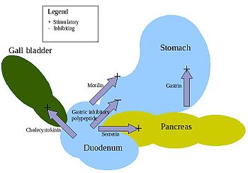
Action of the major digestive hormones
There are at least five hormones that aid and regulate the digestive
system in mammals. There are variations across the vertebrates, as for
instance in birds. Arrangements are complex and additional details are
regularly discovered. For instance, more connections to metabolic
control (largely the glucose-insulin system) have been uncovered in
recent years.
- Gastrin - is in the stomach and stimulates the gastric glands to secrete pepsinogen(an inactive form of the enzyme pepsin) and hydrochloric acid. Secretion of gastrin is stimulated by food arriving in stomach. The secretion is inhibited by low pH .
- Secretin - is in the duodenum and signals the secretion of sodium bicarbonate in the pancreas and it stimulates the bile secretion in the liver. This hormone responds to the acidity of the chyme.
- Cholecystokinin
(CCK) - is in the duodenum and stimulates the release of digestive
enzymes in the pancreas and stimulates the emptying of bile in the gall bladder. This hormone is secreted in response to fat in chyme.
- Gastric inhibitory peptide
(GIP) - is in the duodenum and decreases the stomach churning in turn
slowing the emptying in the stomach. Another function is to induce insulin secretion.
- Motilin - is in the duodenum and increases the migrating myoelectric complexpepsin.
Significance of pH in digestion
Digestion is a complex process which is controlled by several factors. pH
plays a crucial role in a normally functioning digestive tract. In the
mouth, pharynx, and esophagus, pH is typically about 6.8, very weakly
acidic. Saliva controls pH in this region of the digestive tract. Salivary amylase is contained in saliva and starts the breakdown of carbohydrates into monosaccharides.
Most digestive enzymes are sensitive to pH and will not function in a
low-pH environment like the stomach. A pH below 7 indicates an acid,
while a pH above 7 indicates a base; the concentration of the acid or base, however, does also play a role.
The pH of the stomach is very low (highly acidic) which inhibits the breakdown of carbohydrates while there. The strong acid content of the stomach provides two benefits; it serves to denature proteins for further digestion in the small intestines, and provides non-specific immunity, retarding or eliminating various pathogens.
In the small intestines, the duodenum provides critical pH balancing
to activate digestive enzymes. The liver secretes bile into the
duodenum to neutralise the acidic conditions from the stomach. Also the
pancreatic duct empties into the duodenum, adding bicarbonate to neutralize the acidic chyme, thus creating a neutral environment. The mucosal tissue of the small intestines is alkaline with a pH of about 8.5.
Uses of animal gut by humans
- The stomachs of calves have commonly been used as a source of rennet for making cheese.
- The use of animal gut strings by musicians can be traced back to the third dynasty of Egypt. In the recent past, strings were made out of lamb gut. With the advent of the modern era, musicians have tended to use strings made of silk, or synthetic materials such as nylon or steel.
Some instrumentalists, however, still use gut strings in order to evoke
the older tone quality. Although such strings were commonly referred to
as "catgut" strings, cats were never used as a source for gut strings[citation needed].
- Sheep gut was the original source for natural gut string used in racquets, such as for tennis. Today, synthetic strings are much more common, but the best gut strings are now made out of cow gut.
- Gut cord has also been used to produce strings for the snares which provide the snare drum's characteristic buzzing timbre. While the snare drum currently almost always uses metal wire rather than gut cord, the North African bendir frame drum still uses gut for this purpose.
- "Natural" sausage hulls (or casings) are made of animal gut, especially hog, beef, and lamb. Similarly, Haggis is traditionally boiled in, and served in, a sheep stomach.
- Chitterlings, a kind of food, consist of thoroughly washed pig's gut.
- Animal gut was used to make the cord lines in longcase clocks and for fuseebracket clocks, but may be replaced by metal wire.
- The oldest known condoms, from 1640 AD, were made from animal intestine.
|


 Thebusinessalert.com